
Small Intestine Histology Histology slides, Human anatomy and physiology, Physiology
Course Small intestine (anterior view) This seven meter long tube, comprising the duodenum, jejunum and ileum, is the longest portion of the alimentary canal. The small intestine commences its convoluted course through the abdomen at the wider pyloroduodenal sphincter and terminate at the more narrow ileocecal valve.

Small Intestine Section Under The Microscope Stock Photo Download Image Now Ileum, Jejunum
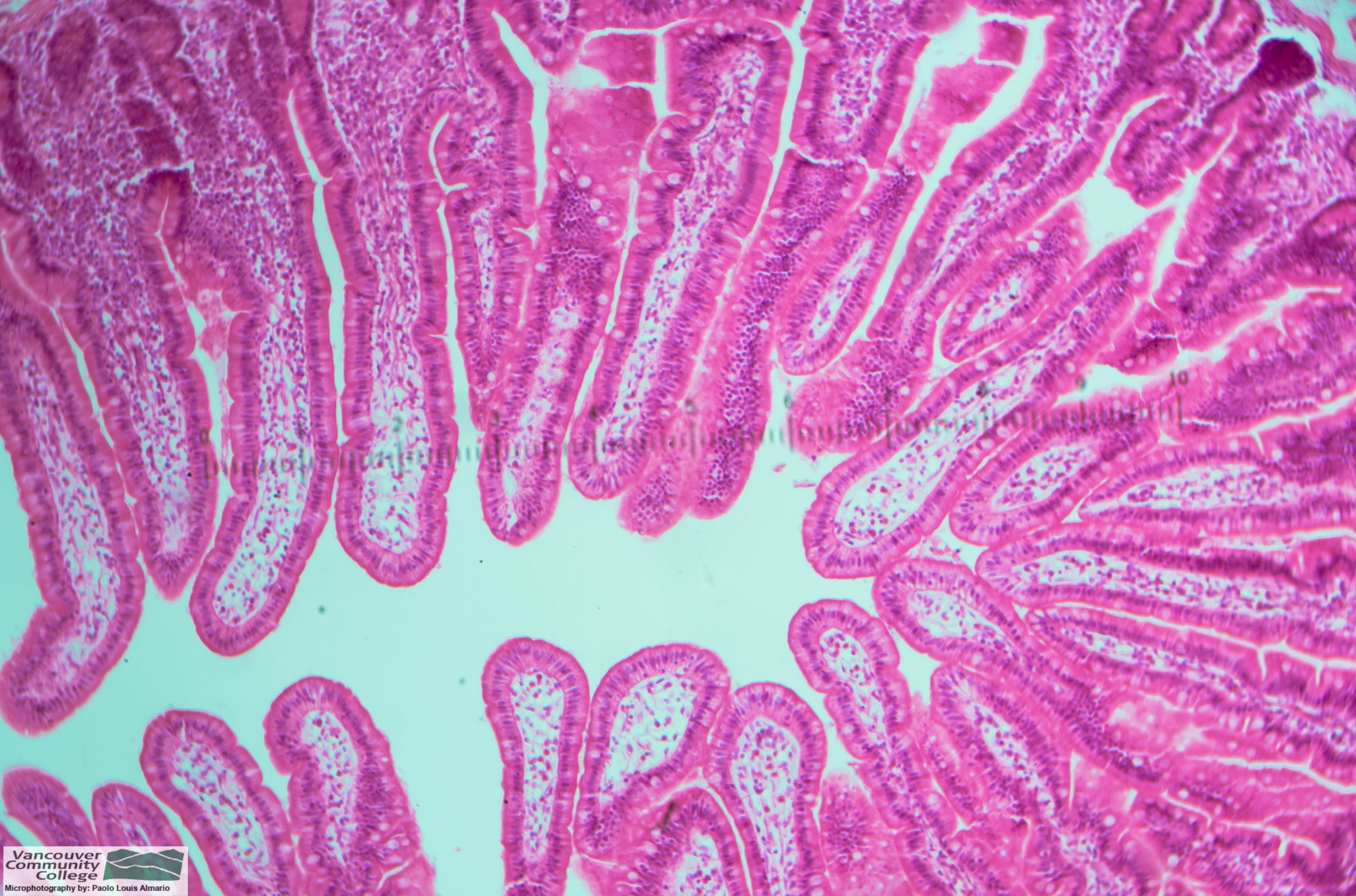
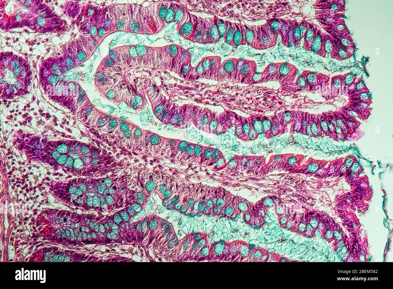
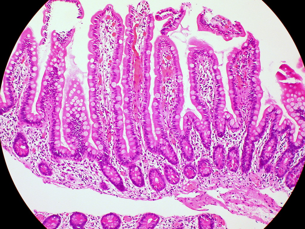
This virtual slide of the SMALL INTESTINE is a cross section of the ileum. In this slide be sure you can identify the following: Simple columnar epithelium (with goblet cells): The epithelium covers finger-like villi (singular: villus) projecting into the opening of the intestine, each with a lacteal (lymph capillary) in its center.
Small Intestine (COLUMNAR EPITHELIUM) LPO MAG by Spyrogenes on DeviantArt
Goblet cells are intestinal mucosal epithelial cells that serve as the primary site for nutrient digestion and mucosal absorption. [2] The primary function of goblet cells is to synthesize and secrete mucus. [1]
Small intestine with villi under the microscope 200x Stock Photo Alamy
Gross description. Small intestine is approximately 6 - 7 m in length ( Gastrointest Endosc Clin N Am 2017;27:1 ) Duodenum: Retroperitoneal, except for first part. Common bile duct and pancreatic duct enter the second part of the duodenum at the ampulla of Vater. Suspensory duodenal ligament (ligament of Treitz) divides duodenum from jejunum.

Small Intestine with Villi Under the Microscope Stock Image Image of macro, research 195449937
The duodenum is the first of the three parts of the small intestine that receives partially digested food from the stomach and begins with the absorption of nutrients. It is directly attached to the pylorus of the stomach.

Simple Columnar Epithelium Small Intestine Supporting Connective Tissu HighRes Stock Photo
A. Simple columnar epithelium. Slide 29 (small intestine) View Virtual Slide Slide 176 40x (colon, H&E) View Virtual Slide Remember that epithelia line or cover surfaces. In slide 29 and slide 176, this type of epithelium lines the luminal (mucosal) surface of the small and large intestines, respectively. Refer to the diagram at the end of this chapter for the tissue orientation and consult.

Distill Confuse chant ileum histology labeled Outflow Brown mud
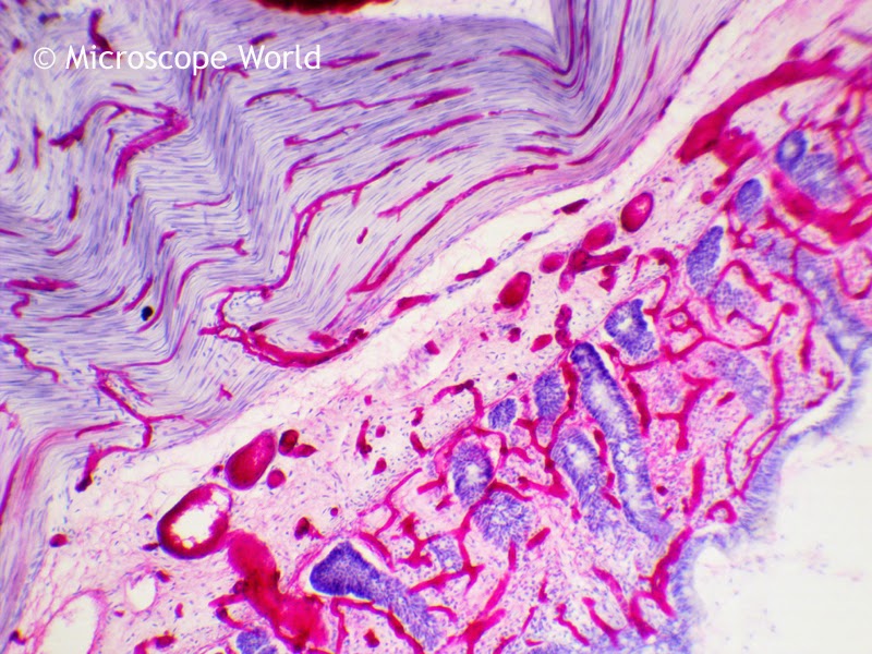
The Small Intestine's Layers. Section of duodenum: This image shows the layers of the duodenum: the serosa, muscularis, submucosa, and mucosa. The small intestine has four tissue layers: The serosa is the outermost layer of the intestine. The serosa is a smooth membrane consisting of a thin layer of cells that secrete serous fluid, and a thin.
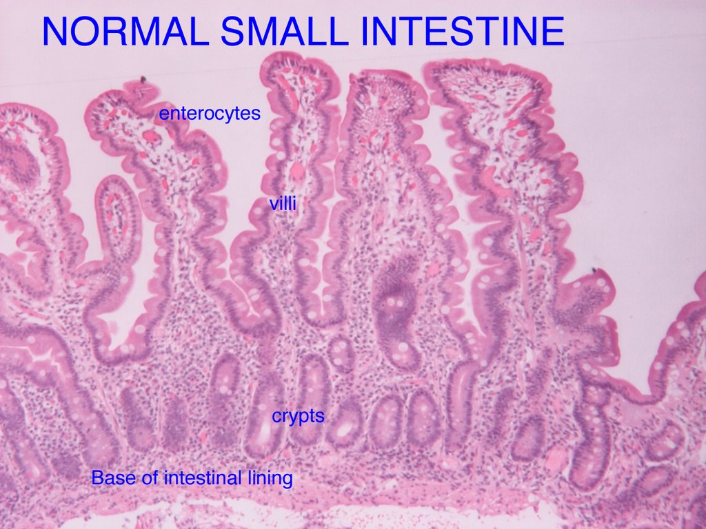
The Food and Gut Journal What does the normal small intestine look like under the microscope?
The histopathological examination of the small intestine under the light microscope: A, control group; B, Cisplatin treated group; c: congestion, d: degeneration, e.

Small intestine Digestive Function, Structure & Length Britannica
You are looking at part of the small intestine. The inner surface of the intestinal wall is made of simple columnar epithelium (sce). Instead of being smooth, the inside of the intestine is folded and covered by millions of tiny projections called villi.

Histology Small Intestine Ileum MM = muscularis mucosae SM= submucosa V = villi ME
Small intestine cancer happens when malignant () cells form in your small intestine, or small bowel. Your small intestine is part of your body's digestive system, which includes organs like your liver, pancreas gallbladder, as well as your gastrointestinal (GI) tract.

Histology Small intestine Medical Laboratory, Medical Science, Medical School, Tissue Biology
The small intestine is 4-6 metres long in humans. To aid in digestion and absorption: the small intestine secretes enzymes and has mucous producing glands. The pancreas and liver also deliver their exocrine secretions into the duodenum. The mucosa is highly folded. large circular folds called plicae circulares (shown in the diagram to the right.
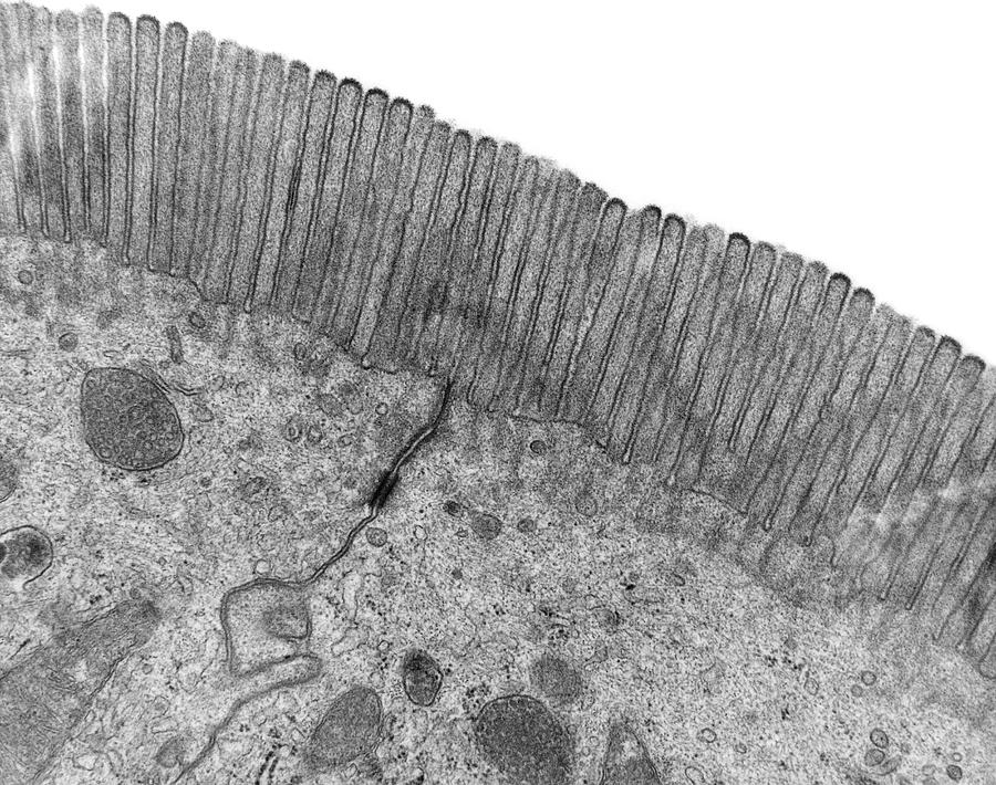
Microvilli Of The Small Intestine Photograph by Dennis Kunkel Microscopy/science Photo Library
The small intestine is the site where digestion is completed, using enzymes from the pancreas and bile, and where products of digestion are absorbed. The small intestine possesses several features that increase the surface area for digestion and absorption, including its long length, the presence of plicae circulares, numerous villi, and the simple columnar epithelium with microvilli (brush.
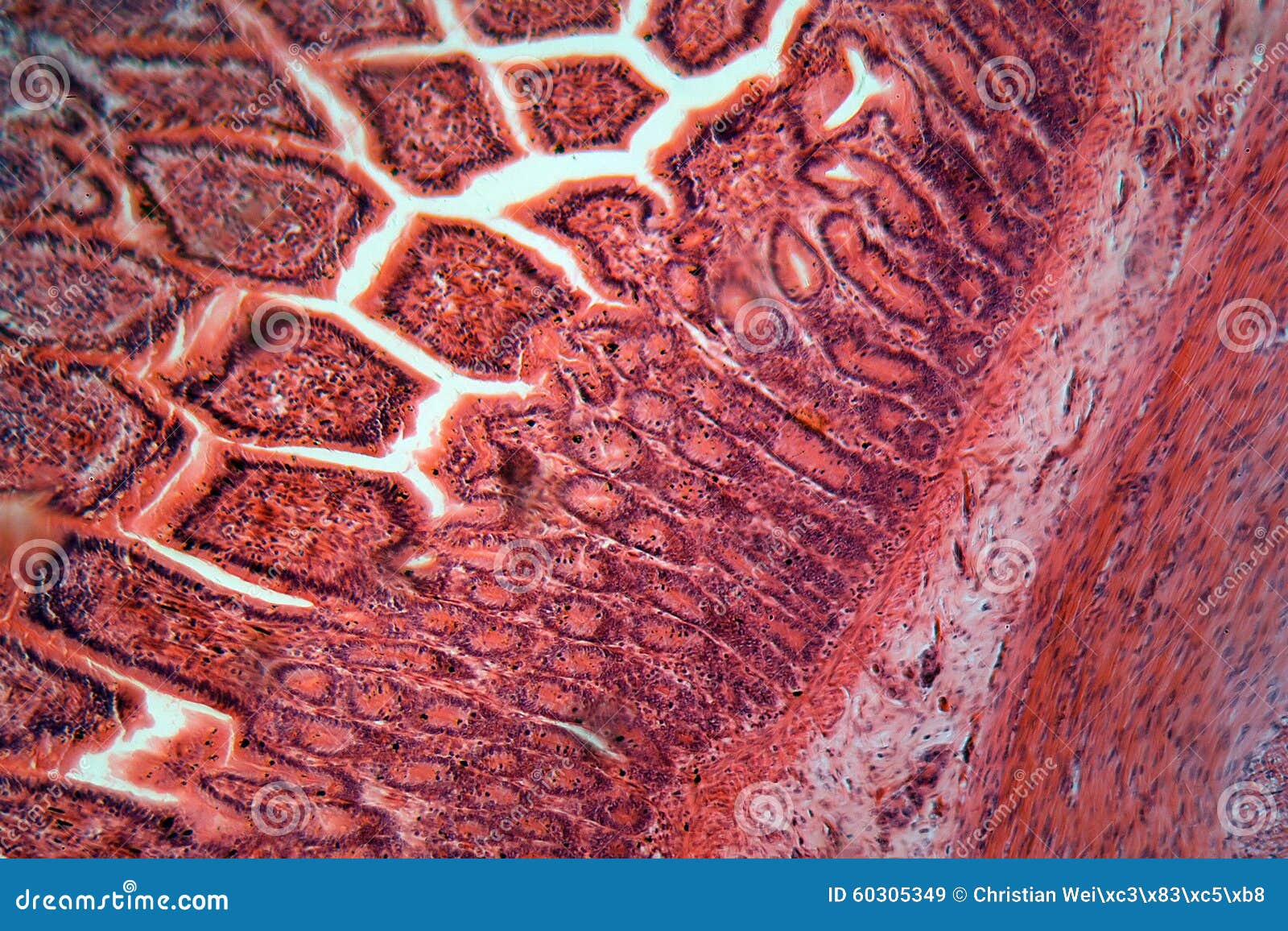
Intestine Cells Under the Microscope Stock Image Image of micrograph, laboratory 60305349
The histology of the wall of the small intestine differs somewhat in the duodenum, jejunum, and ileum, but the changes occur gradually from one end of the intestine to the other. 1. Duodenum. Slide 162 40x (pyloro-duodenal junct, H&E) View Virtual Slide. Slide 161 40x (pylorus, duodenum, pancreas, H&E) View Virtual Slide. Look at slide 162 first.
Normal Small Intestine Mucosa Flickr Photo Sharing!
A bulk of the small intestine is suspended from the body wall by an extension of the peritoneum called the mesentery. As seen in the image to the right, blood vessels to and from the intestine lie between the two sheets of the mesentery. Lymphatic vessels are also present, but are not easy to discern grossly in normal specimens.

Representative light microscopic histological view of intestinal villi... Download Scientific
It is thus crucial to dissect key differences in both ecology and physiology between small and large intestine to better leverage the immense potential of human gut microbiota imprinting, including probiotic engraftment at biological sensible niches.
Microscope World Blog Small Intestine Microscopy Images
Staining is widely used in histopathology and diagnosis, as it allows for the identification of abnormalities in cell count and structure under the microscope. A huge range of stains is used in histology, from dyes and metals to labeled antibodies.